
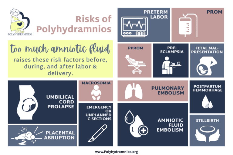
Risks of Polyhydramnios
Polyhydramnios raises your risks of several pregnancy complications including preterm labor, premature rupture of membranes, preterm premature rupture of membranes, pre-eclampsia, fetal malpresentation, umbilical cord prolapse, placental abruption, emergency and unplanned c-sections, stillbirth, post-partum hemorrhage, macrosomia, pulmonary embolism, and amniotic fluid embolism. As your fluid level increases, so do your risks of experiencing one or more of these complications.
Table of Contents
Risks
Preterm Labor
Generally, pregnancy lasts an average of 40 weeks to allow for optimum development of the fetus. Labor that happens from 37 weeks and beyond is usually allowed to progress without intervention as 37 weeks is considered “early term” and most babies born after this week do just fine without assistance. Preterm labor is labor that begins before 37 weeks gestation. A baby born before 37 weeks is called “premature”; premature babies statistically have more health problems both as babies and later in life than babies born at term.
Polyhydramnios can increase your risk for preterm labor for several reasons:
- The strain that the extra fluid puts on your uterus can cause an irritable uterus. 18% of women with an irritable uterus go into preterm labor.
- Many factors that cause Polyhydramnios are also risk factors for preterm labor including, diabetes, infections, and certain birth defects in your baby.
- Polyhydramnios can lead to preeclampsia, which is also a risk factor for preterm labor.
If you go into preterm labor, your healthcare provider may give you medications to try to stop it. They may also give your baby steroids to help with their lung development if premature birth seems likely. The symptoms of preterm labor include changes in vaginal discharge, pressure low in your pelvis, constant low, dull backache, belly cramps, regular or frequent contractions, or your water breaks. If you have signs of preterm labor, notify your healthcare provider.
Premature Rupture of Membranes (PROM)
Premature rupture of membranes is when the amniotic sac (aka: your water) breaks before labor begins, but after 37 weeks so baby is considered to be at term. PROM is concerning because it can lead to infections, placental abruption, cord compression, c-section, and postpartum infection. Two possible causes of PROM are a natural weakening of the membranes and the force of contractions, both of which are exaggerated by Polyhydramnios, therefore increasing the risk for PROM.
The signs of PROM may include having a big gush of fluid, a slow steady leaking, or a feeling of constant wetness. PROM can be detected by cervical exam, testing the fluid ph, looking for ferning pattern of fluid under a microscope, and measurement of fluid levels using ultrasound. If PROM is detected, there are certain medical protocols in place to decide how to proceed and might include antibiotics for infections, corticosteroids for fetal lung development, and delivery of your baby.
Preterm Premature Rupture of Membranes (PPROM)
The only difference between PROM and PPROM, is that PPROM is when your water breaks before 37 weeks. It is often caused by an infection in the uterus.
Management for PPROM is highly dependent on your circumstances and the gestational age in which it presents. Most cases of PPROM are treated by hospitalization, expectant management, monitoring, corticosteroids for fetal lung development, antibiotics for infection, and tocolytics to stop preterm labor. The goal should be to stabilize mom and deliver at 34 weeks. If that is not possible, earlier delivery may be necessary.
Pre-eclampsia*
Preeclampsia is a serious pregnancy-related disorder characterized most often by high blood pressure and protein in the urine. Although recent guidelines from the ACOG have stated that the diagnosis can be made without the presentation of protein in urine if other clinical indications are present. Complications of preeclampsia can include maternal brain injury, impaired kidney and liver function, blood clotting problems, pulmonary edema, seizures, or in severe untreated cases, infant or maternal death. The exact causes of preeclampsia are still unknown but many researchers agree that it seems to have a link to problems with the placenta.
*As we researched for Polyhydramnios.org, we couldn’t find any clear evidence that Polyhydramnios raises the risk of preeclampsia. We did however find several sources that state there is a link and we have seen first hand evidence of preeclampsia in women with Polyhydramnios, so we included it here as a possible risk factor of Polyhydramnios.
Fetal Malpresentation
The ideal position of your baby for a vaginal delivery is head down pressing on your cervix, facing your back, with its chin tucked to its chest (vertex position). The majority of babies naturally move into this position sometime before labor begins. When a baby is in any other position it is called fetal malpresentation.
There are many types of fetal malpresentation, these are the most common:
- Breech is when the baby is positioned with its butt or feet pressing against your cervix.
- Transverse is when baby is lying sideways so that its back, shoulders, arms, or legs could enter the birth canal first.
- Oblique is when the baby is up high with its head is pointing toward your hip so no part is against your cervix.
- Unstable lie is when baby keeps flipping positions with no parts pressing against your cervix.
- Funic or cord presentation is when the umbilical cord is pressing against your cervix.
Malpresentation can increase the risk of PROM, uterine rupture, cord prolapse, assisted delivery, c-section, and birth injuries to both you and your baby. Malpresentation occurs in about 4% of normal pregnancies, but your risk is higher with Polyhydramnios because the extra amniotic fluid gives your baby more room to move around.
If you are nearing labor and your baby is in an undesirable position, your healthcare provider may suggest options for attempting to turn your baby. External Cephalic Version (ECV) is a procedure done in the hospital where your healthcare team attempts to manipulate your baby from the outside to get it to turn. The success rate for ECV is approximately 58% and it carries more risks for moms with Polyhydramnios. Some mommas have also had success getting their babies to turn using acupuncture or Spinning Babies techniques, although these techniques are not backed by reliable evidence.
*Never attempt any inverted (upside down) Spinning Babies positions with Polyhydramnios since this can increase your risk of cord prolapse.
Umbilical Cord Prolapse
Umbilical cord prolapse is when the umbilical cord slips into the birth canal either before or alongside of your baby’s presenting part. This is a medical emergency because it puts the umbilical cord in position to become compressed between your baby and your bony pelvis, depriving your baby of the oxygen and nutrients it depends on. A compressed cord causes carbon monoxide to quickly build up in the baby’s blood stream leading to respiratory acidosis. Cord compression can also lead to brain damage, fetal hypoxia (oxygen deprivation), and even death.
When umbilical cord prolapse is diagnosed delivery should quickly follow. Emergency c-section is the usual method of delivery except in rare cases where a vaginal or assisted delivery is deemed faster. The best outcomes happen when the time interval between diagnosis and delivery is less than 30 minutes. This is why your baby has a better prognosis if cord prolapse happens in the hospital (3% mortality rate) as opposed to at home (44% mortality rate).
If prolonged time until delivery is anticipated, one of these methods should be used to help relieve compression of the cord until delivery can take place:
- Manual elevation of the fetal presenting part using either two fingers or the entire hand through the vagina.
- Positioning the patient in steep Trendelenburg, exaggerated Sim’s position, or knee-chest position.
- Filling the bladder with 500-700cc or more of saline.
- Tocolytics may help, but only with careful consideration because they may cause uterine atony after delivery.
- Cord replacement (NOT recommended unless very specific criteria can be met).
Polyhydramnios is a risk factor for umbilical cord prolapse because excess amniotic fluid leads to fetal malpresentation, the fetus not engaging in the pelvis before labor, and PROM. All of these things are linked to higher incidence of umbilical cord prolapse because in each of these situations, there is free space between the presenting part of your baby and the opening of the cervix allowing the cord a chance to slip through and become compressed.
The risk of umbilical cord prolapse is a contributing factor to why many women with Polyhydramnios are induced. Here are some things to consider:
- Unstable lie after 37 weeks is an indication for 38-39 week elective c-section as a preventative measure. If a vaginal delivery is desired, ECV may be attempted with immediate induction to follow and controlled rupture of membranes.
- Caution should be exercised in patients at risk for umbilical cord prolapse during AROM (artificial rupture of membranes). AROM should not be done before the head is well applied to the cervix unless necessary.
- Controlled rupture by an experienced obstetrician is recommended using a hypodermic needle or pudendal block trumpet to achieve a slow and controlled drain of amniotic fluid.
- When umbilical cord prolapse happens, the outcome is considerably more favorable when each of these factors also occur:
- Prolapse happens inside of the hospital.
- Time from diagnosis to delivery is less than 30 minutes.
- Birth weight is greater than 5.5 pounds (2500 grams).
- A c-section delivery is performed.
Placental Abruption
During pregnancy, your placenta forms and attaches itself to the lining of your uterine wall. It’s main function is to act as the mediator between you and your baby, providing your baby with nutrients and oxygen and also filtering waste from your baby’s blood. After your baby is delivered, the placenta is no longer needed, so your body will expel it from your uterus. Placental abruption is when this process starts to happen before your baby is born. The risk for placental abruption is higher with Polyhydramnios because of the increased risk of PROM or having a leak in your amniotic sac. Both of these incidences can make your placenta tear away early.
Placental abruption can happen little by little, slowly tearing away from the uterine wall, but it usually happens quite suddenly. There is no way to know for sure if you’ve had a placental abruption until the placenta has been delivered and examined, so your health care provider will diagnose placental abruption based off of your clinical symptoms. Signs of a placental abruption may include vaginal bleeding, back pain, painful or tender belly, repeated contractions, or problems with the baby’s heart rate.
If a minor abruption is suspected it may not cause any problems, and your healthcare provider may choose to monitor you more closely. If a major abruption is suspected, however, immediate delivery may need to take place to prevent complications such as major bleeding, blood clots, organ failure, maternal death, or stillbirth. A major placental abruption is a catastrophic event and requires immediate attention.
Emergency or Unplanned C-Section
A cesarean section (c-section) is a major operation where incisions are made through the abdominal and uterine walls to deliver your baby and the placenta. Sometimes a c-section is planned ahead of time, but sometimes complications arise and quick action is needed. In these cases, an emergency or unplanned c-section will likely take place.
An unplanned c-section is one that usually happens because labor is not going as well as expected. An unplanned c-section is urgent, but not usually due to a life-threatening reason. The time from decision to do a c-section to delivery may be anywhere from 30 minutes to an hour, and you will likely be awake during the procedure. Reasons for an unplanned c-section may include stalled labor, weak contractions, baby not tolerating labor, or fetal malpresentation.
An emergency c-section is one that happens very quickly due to a life-threatening complication for either you or your baby. The time from the beginning of surgery to delivery in this case can be as little as one minute! In some cases there may be time and opportunity for epidural medication, but if there is not, general anesthesia will be used and you will be asleep for the procedure. Reasons for an emergency c-section include fetal or maternal distress, umbilical cord prolapse, hemorrhage, placental abruption, and uterine rupture.
An emergency c-section increases the risk of severe hemorrhage, complications from anesthesia, and accidental injury to you or your baby.
Polyhydramnios increases the risk of an emergency or unplanned c-section because it also increases the risk of stalled labor, fetal anomalies that may make it hard for baby to tolerate labor, fetal malpresentation, cord prolapse, hemorrhage, and placental abruption.
Stillbirth
Stillbirth is when your baby passes away while still in the womb but after 20 weeks gestation. The reason that stillbirth occurs is still widely unknown, but medical experts have identified several risk factors that make it more likely to happen. Since Polyhydramnios puts you at risk for some of these factors, you are also at an increased risk of stillbirth. These risk factors include infection, placental abruption, cord prolapse, preeclampsia, preterm labor, birth defects, genetic conditions, and rh disease.
Postpartum Hemorrhage
Postpartum hemorrhage is when you have excessive bleeding after the delivery of your baby which can cause a dangerous drop in your blood pressure resulting in shock or death. It usually happens immediately following delivery, but it can happen later in the postpartum period. Postpartum hemorrhage happens either when your uterus doesn’t contract properly after delivery, or when small pieces of the placenta remain inside the uterus after the rest has been expelled. There are many risk factors for postpartum hemorrhage and Polyhydramnios raises your risk for some of them including having a placental abruption, an overdistended uterus, preeclampsia, infection, medications to induce labor, medications to stop preterm labor contractions, and general anesthesia.
The signs of postpartum hemorrhage may include uncontrolled bleeding, drop in blood pressure, elevated heart rate, and decrease in red blood cell count. After delivery, your healthcare team will check for signs of uterine atony (when your uterus remains soft after delivery) since this is a major sign that it is not contracting properly. Your healthcare team will also monitor your blood loss by counting saturated pads or by weighing packs and sponges used to absorb blood.
Treatment for postpartum hemorrhage depends on the cause and severity of the hemorrhage and may include medication to stimulate contractions, uterine massage, removal of pieces of the placenta still inside the uterus, surgical or instrumental methods to compress bleeding inside the uterus after a c-section, laparotomy, and hysterectomy. It may also be necessary to replace fluids and blood intravenously, and to provide oxygen using a mask in order to prevent shock.
Macrosomia
Macrosomia literally means “large body” and it is the term used to describe a baby born weighing more than 8 pounds 13 ounces (4,000 grams), regardless of their gestational age. Fetal macrosomia can only be diagnosed once your baby has been born and weighed, although there are ways that your healthcare provider will use to estimate if they think your baby will have macrosomia. The biggest Polyhydramnios related risk factors for macrosomia include having maternal diabetes or if other maternal risk factors are ruled out, certain medical conditions in your baby.
Macrosomia carries risks such as shoulder dystocia, birth injuries to you or your baby, needing an assisted vaginal delivery, c-section, postpartum hemorrhage, uterine rupture (if you’ve had a previous uterine surgery), low blood sugar in your baby, and childhood obesity or metabolic syndrome in your baby later on in their childhood.
Pulmonary Embolism
A pulmonary embolism (PE) is a life-threatening emergency where a blood clot blocks an artery in your lung. Postpartum pulmonary embolism can occur during pregnancy, but most often happens soon after labor and delivery. Pregnancy and the postpartum period in general increase a woman’s risk of PE but Polyhdyramnios can also increase your risk if there is excessive weight gain, a period of bed rest or low activity, or c-section delivery.
The symptoms of PE most often include shortness of breath, chest pain, and leg swelling. If you notice these symptoms after delivery (or up to 4 weeks postpartum), alert your healthcare provider immediately. If PE is suspected, it can be diagnosed by blood tests and diagnostic imaging. PEs are treated with blood thinners, and in emergency situations, thrombolytics are used to break up the blood clot.
There are things you can do to try to help prevent a blood clot from forming. These include staying active while pregnant and maintaining a healthy weight, requesting to wear compression socks during a c-section, and getting up to move early and often after delivery.
Amniotic Fluid Embolism
Amniotic fluid embolism (AFE) is an extremely rare birth complication characterized by a two-phase response of an allergic-like reaction to amniotic fluid entering your circulatory system. In phase one, you will experience rapid respiratory failure which can cause cardiac arrest. In phase two, hemorrhaging begins and leads to DIC, which prevents your blood from clotting, and requires a blood transfusion. Even though AFE is very rare, it is a serious complication and one of the leading causes of maternal death worldwide. It is estimated that up to 50% of mothers who die from AFE do so within an hour of the onset of their symptoms.
The exact cause of AFE is unknown. Studies have shown that some women have fetal matter enter their circulatory system and experience no problems, while others have deadly reactions. Although the exact cause is unknown, Polyhydramnios has been listed as a risk factor due to the excess amount of amniotic fluid in the uterus.
AFE usually happens during labor, birth, or immediately postpartum. Some of the signs of AFE include increased anxiety, agitation, impending sense of doom, confusion, nausea, vomiting, chills, skin discoloration, shortness of breath, fetal distress, and abnormal vitals. These symptoms quickly lead to loss of consciousness, seizure or cardiac arrest, and hemorrhage. There is no way to prevent or predict AFE. There is also no cure for AFE, only supportive care to try to quickly manage the symptoms.
FAQs
FAQs
Polyhydramnios is a secondary condition, meaning it is usually caused by another underlying condition. So, in short, no, it does not cause birth defects. It can, however, sometimes be the result of a birth defect in your baby. Read more about the causes of Polyhydramnios.
Treatment for Polyhydramnios involves effective management of the symptoms and the underlying causes. Management of the underlying cause varies widely and may include anything from diet changes to in-utero surgery. Some symptoms can be managed by rest, the elevation of swollen limbs, hydration, and sleeping in a propped-up position. If Polyhydramnios is very severe and breathing is impaired, amnioreduction may provide temporary relief. Read more about managing polyhydramnios here.
Read Next
Causes of Polyhydramnios – Nearly half of Polyhydramnios cases happen for unknown reasons, but after idiopathic cases, the leading causes of polyhydramnios are genetic anomalies in babies.